Protein A/G Magnetic Beads are a critical tool in modern molecular biology and immunology laboratories, frequently used for antibody purification, immunoprecipitation, and other immunoassay applications. These beads leverage the binding capabilities of both Protein A and Protein G, which provide optimal binding to a wide range of antibodies from various species, enhancing their versatility and application across diverse experimental needs.
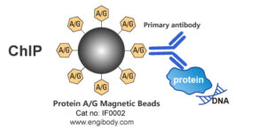
Structure and Binding Specificity
Protein A/G is a recombinant fusion protein that combines Protein A and Protein G domains, allowing it to bind to the Fc region of immunoglobulins (IgG) from several species. This makes Protein A/G beads highly adaptable, as they can be used with antibodies that either Protein A or Protein G alone might not effectively bind. The details of how Protein A and Protein G interact with different immunoglobulins are well-documented on resources like the National Institutes of Health (NIH) and in educational resources at Harvard University.
Applications in Antibody Purification
One of the primary uses of Protein A/G Magnetic Beads is in antibody purification. By attaching antibodies from complex samples to the beads, researchers can isolate and concentrate antibodies with high specificity and efficiency. This process is particularly valuable in therapeutic antibody production, where purity is essential. Detailed protocols for antibody purification with Protein A/G beads can be found in resources like the National Center for Biotechnology Information (NCBI) and University of California, San Diego (UCSD).
Immunoprecipitation and Co-Immunoprecipitation
Protein A/G Magnetic Beads are extensively used in immunoprecipitation (IP) and co-immunoprecipitation (Co-IP) experiments. In these applications, antibodies bound to the beads capture their target antigens from a mixture, enabling analysis of protein-protein interactions or detection of specific proteins in complex samples. The Centers for Disease Control and Prevention (CDC) provides information on protein interactions and their roles in disease pathways. Additionally, educational guides on Co-IP techniques are available at Stanford University.

Magnetic Properties and Easy Handling
The magnetic nature of Protein A/G beads enables easy separation from the sample matrix. When exposed to a magnetic field, the beads quickly localize, simplifying washing steps and reducing sample loss. This feature makes them ideal for high-throughput applications, as discussed in technical resources from MIT and the National Institutes of Standards and Technology (NIST).
Protein A/G Beads vs. Protein A or Protein G Beads Alone
Choosing between Protein A/G, Protein A, or Protein G depends on the antibody subclass and species. While Protein A or Protein G beads may be specific for certain species or subclasses, Protein A/G beads offer broader binding capabilities. For an in-depth comparison, you can refer to studies available from National Library of Medicine (NLM) and the U.S. Food and Drug Administration (FDA).
Common Protocols and Guidelines
Standard protocols for using Protein A/G Magnetic Beads in various applications can be found in scientific literature and online databases such as PubMed and the U.S. National Institute of Allergy and Infectious Diseases (NIAID). Additionally, institutions like Yale University provide online lab manuals detailing specific usage and troubleshooting tips for Protein A/G beads.
Optimization Tips for Protein A/G Beads
Optimizing binding conditions for Protein A/G Magnetic Beads can enhance the yield and specificity of antibodies. For example, pH adjustments and buffer modifications play a significant role, as discussed in academic research available from University of California, San Francisco (UCSF) and Johns Hopkins University. Additionally, the use of different elution buffers can help minimize contamination, with detailed protocols available from the National Institute of Environmental Health Sciences (NIEHS).
Advanced Applications in Clinical and Research Settings
Protein A/G Magnetic Beads are also used in advanced applications, such as isolating biomarkers from serum for early disease detection or in the development of new diagnostic tools, as shown in studies by National Cancer Institute (NCI) and the U.S. Department of Energy (DOE). For example, these beads are utilized in identifying potential biomarkers for autoimmune disorders, cancer, and infectious diseases.
Concluding Remarks
Protein A/G Magnetic Beads provide a versatile and efficient solution for antibody-based applications, including immunoprecipitation, antibody purification, and biomarker research. For more extensive protocols and additional technical information, consider visiting educational sites like Columbia University and government resources such as the National Institute on Aging (NIA). The use of these beads in both research and clinical settings underscores their significant role in advancing molecular biology and biomedical research.